
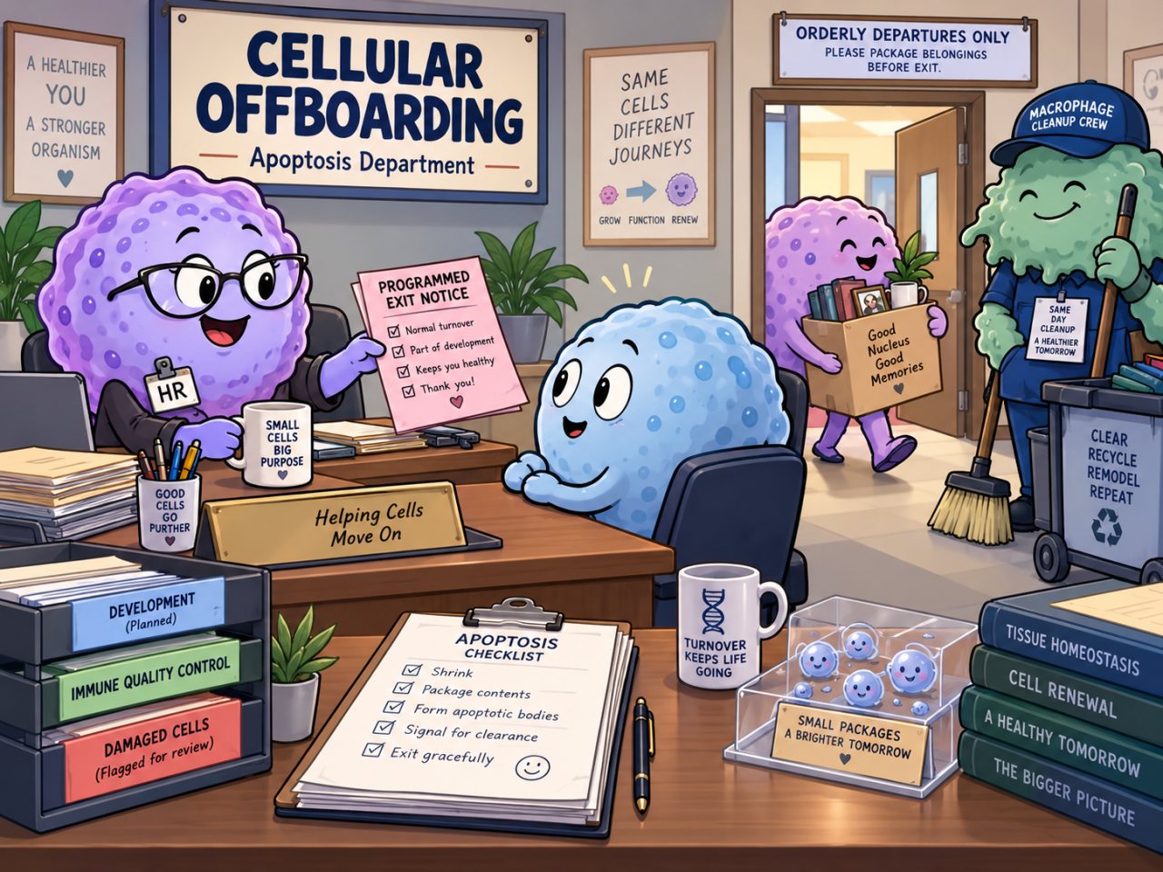
Your cells have a built-in self-destruct mechanism.
Really!
It's a process called apoptosis, which is also a very fun word to say aloud.
A... POP... TOE... SIS....
Cells actually have genes in their DNA that help control the process of apoptosis, leading to the orderly death of the cell. Some of these pro-apoptotic genes have even been nicknamed "suicide genes." If you need a refresher on what genes are, see the Confidential Genetic Files Never Leave Headquarters! scandal.
And this "programmed cell death" is actually an important part of both development and the ongoing maintenance of your body's tissues.
For this article, we're focusing on the surprisingly routine version of cellular self-destruction.
Normal cells.
Dying at a normal rate.
For perfectly normal reasons.
Biology is reassuring like that.
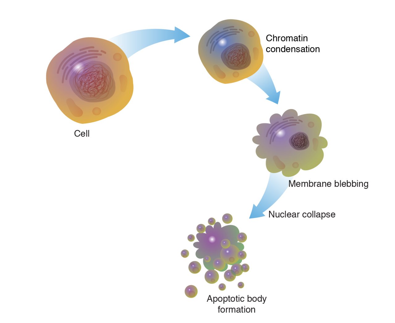
When a cell activates its suicide pathway, it sets off a molecular demolition crew. Proteins called caspases become activated and begin cutting up other proteins inside the cell, helping dismantle it from within. The cell shrinks and breaks its contents into tidy membrane-bound packages. Then the body's cellular janitors, immune cells called macrophages, come by and sweep up the remains.
Very civilized.
And apparently the body uses this offboarding process all over the place.
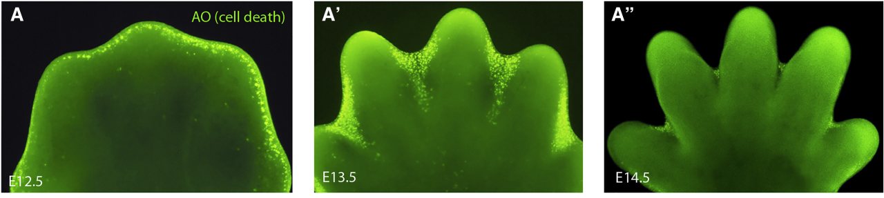
During development, for example, your early hands are considerably less hand-shaped than the finished product. Cells between the developing fingers are informed that their services will no longer be required. Apoptosis helps remove them, leaving you with individual fingers instead of permanent little paddles.
The immune system is even more ruthless about personnel decisions. It produces enormous numbers of immune cells, tests them, and eliminates many that are unnecessary, defective, or potentially dangerous.
IMMUNE SYSTEM: Thank you for your interest in defending the body. Unfortunately, we have decided to proceed with other immune cells at this time. Initiate self-destruct protocol in 10… 9… 8…
And sometimes a cell becomes sufficiently damaged that continuing to employ it would pose a risk to the entire organization. Ideally, apoptosis hands it a cardboard box, supervises the cleaning out of its desk, and escorts it from the building before it can become everybody else's problem.
Because when cells that should die start refusing to leave?
Well… That's another scandal.
The Real Story
Apoptosis is a highly controlled form of programmed cell death. Instead of bursting open and spilling its contents everywhere, an apoptotic cell dismantles itself in an organized way.
Proteins called caspases play a major role in that process. Once activated, they begin cutting apart other proteins inside the cell and help shut down its normal machinery. The cell shrinks, its DNA is broken into pieces, and its outer membrane begins forming small bulges before the cell separates into tidy membrane-bound packages called apoptotic bodies.
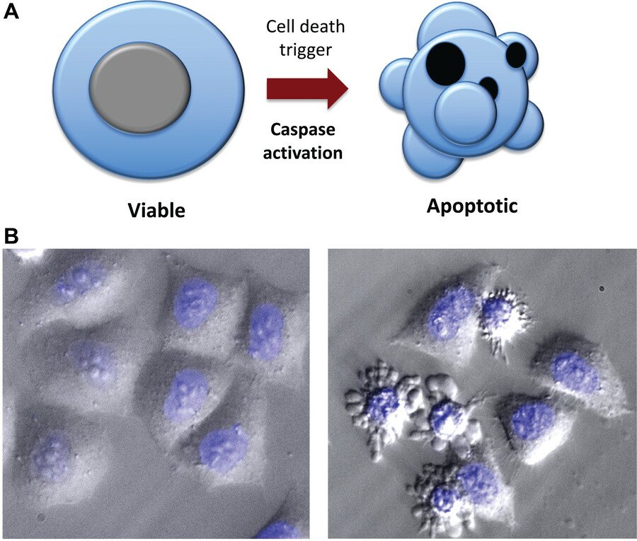
Those packages are then removed by cleanup cells. Some of these are macrophages, immune cells that act like the body’s janitors by surrounding, swallowing, and digesting dead cells and cellular debris. Because the dying cell keeps most of its contents neatly contained until cleanup arrives, apoptosis usually causes much less inflammation than a cell simply rupturing and making a mess.
And this is not just something the body does when a cell is badly damaged. Apoptosis is part of normal life.
One of its most obvious jobs happens during development. Early in development, there is tissue between the forming fingers and toes. Apoptosis helps remove some of those cells as the individual digits take shape.
The immune system also uses apoptosis as part of quality control. Your body produces enormous numbers of immature white blood cells called lymphocytes that are being trained to recognize dangerous targets without attacking your own tissues. Many fail that test. Cells that are unnecessary, malfunctioning, or potentially dangerous can be eliminated through apoptosis before they are allowed to join the active immune system.
Apoptosis also helps keep ordinary tissues from becoming overcrowded. Your body is constantly making replacement cells, especially in tissues that experience a lot of wear and tear. Skin cells are continually replaced as older cells are lost from the surface, and cells lining the stomach and intestines are renewed rapidly because they live in such a chemically and mechanically demanding environment. Programmed cell death helps balance cell production with cell removal so tissues maintain roughly the right number of cells.
The reproductive system provides another everyday example. In the inner lining of the uterus, apoptosis increases during the late part of the menstrual cycle and contributes to the normal turnover and remodeling of that tissue.
Somewhere, a reader has just asked whether blocking apoptosis would eliminate periods.
Biology regrets to report that the system is considerably more complicated than that. 😂 Apoptosis is only one part of menstruation, which also involves hormonal changes, inflammation, changes in blood vessels, and breakdown of the tissue surrounding cells.
Apoptosis can also remove cells that have become seriously damaged or potentially dangerous. If a cell accumulates enough internal damage, the safest outcome for the rest of the tissue may be for that cell to shut itself down and be removed.
The important idea in all this is balance.
Too much cell death can contribute to tissue loss and disease.
Too little cell death can allow cells to survive when they should have been removed.
And when cells that should die start refusing to leave?
That is where another scandal begins.
Sources
-
Apoptosis and Cell Death: Signaling in Health and Diseases
Merheb, E., Zubair, M., & Bokhari, S. R. A. (2026). Apoptosis and Cell Death: Signaling in Health and Diseases. StatPearls Publishing.
Why this source matters: Provides an up-to-date overview of apoptosis as a regulated, caspase-mediated process involved in development, immune regulation, tissue homeostasis, and removal of potentially harmful cells. It also supports the descriptions of cell shrinkage, chromatin condensation, membrane blebbing, and phagocytic clearance.
-
Phagocytosis of Apoptotic Cells in Homeostasis
Arandjelovic, S., & Ravichandran, K. S. (2015). Phagocytosis of apoptotic cells in homeostasis. Nature Immunology, 16(9), 907–917.
Why this source matters: Supports the article’s explanation that large numbers of apoptotic cells are routinely removed by professional and nonprofessional phagocytes and that this clearance is integral to development and normal tissue homeostasis.
-
The Control of Apoptosis in Lymphocyte Selection
Strasser, A., & Bouillet, P. (2003). The control of apoptosis in lymphocyte selection. Immunological Reviews, 193, 82–92.
Why this source matters: Supports the role of apoptosis in eliminating useless or self-reactive developing lymphocytes and in controlling immune-cell populations after activation.
-
9.3 Response to the Signal
Clark, M. A., Douglas, M., & Choi, J. (2018). 9.3 Response to the signal. In Biology 2e. OpenStax, Rice University.
Why this source matters: Provides textbook-level support for apoptosis as normal programmed cell death, especially its roles in embryonic development, T-cell selection, and controlled elimination of potentially harmful cells.
-
Apoptosis in Human Endometrium and Endometriosis
Harada, T., Kaponis, A., Iwabe, T., Taniguchi, F., Makrydimas, G., Sofikitis, N., Paschopoulos, M., Paraskevaidis, E., & Terakawa, N. (2004). Apoptosis in human endometrium and endometriosis. Human Reproduction Update, 10(1), 29–38.
Why this source matters: Supports the article’s menstruation example by describing apoptosis as a normal contributor to endometrial cell turnover, particularly during the late secretory and menstrual phases.