
I saw the headline “Human Brain is Two Organs Fused Together, Study Says,” and my response was, “What, now?”
Then I looked at the actual study and was like, “Oh, okay. They’re talking about early brain development, and how the cells destined to become the hindbrain appear to follow a different genetic developmental pathway from the cells that become the forebrain and midbrain.”
And then I forgot about it.
The next day, the news had apparently spread, and family and friends, knowing that I’m a neuroscientist, started texting.
“Wow! We have two brains fused together. That’s so cool!”
And I had to do a major face palm. 🤦♀️
So this current article I'm writing is doing double duty: explaining the science and explaining how easy it is for science news to become inflated as it moves from research paper to headline.
Which is how you can end up with this progression:
Research paper:
“We found evidence that forebrain/midbrain and hindbrain lineages are developmentally distinct very early in embryogenesis.”
Research interpretation:
“We postulate the brain is a composite organ arising from two lineage-restricted progenitors.”
University press release:
“THE HUMAN BRAIN IS TWO SEPARATE ORGANS.”
News headlines:
“HUMAN BRAIN IS TWO ORGANS FUSED TOGETHER!”
My incoming text messages:
“Wow! Science has discovered we have two brains!”
🤦♀️
The study does present genuinely interesting new developmental evidence that could affect the way scientists study brain development, neurological disease, and how different types of neurons are grown in the laboratory.
But two separate organs squished together?
Absolutely not.
Even one of the researchers himself, Dr. Kyle Loh, said:
“The brain is one organ... But it’s built in two different parts that connect and work together.”
And having distinct developmental pathways does not automatically mean something is really two organs. The heart also develops through more than one embryonic developmental program, yet nobody is walking around talking about having two hearts fused together.
So the broader message is to remember that scientific qualifiers have a nasty habit of disappearing during news coverage:
-
“suggests” becomes “shows”
-
“developmental systems” becomes “organs”
-
“distinct embryonic origins” becomes “two brains”
Here’s a handy general guide to remember when interpreting new science news:
Scientists say: “Our data suggest…”
Headline says: “SCIENTISTS PROVE…”
Scientists say: “May contribute to…”
Headline says: “CAUSES…”
Scientists say: “In mice…”
Headline says: “YOU.”
And you still have the same single brain today that you woke up with yesterday.
We just understand a little better how different parts of it got started. 😊
And yes, I am fully aware that this criticism is coming from a website called Science Scandal. Sensational headlines are literally part of the deal here. The difference is that we tell you that up front, and our rule is to make the real science sound scandalous without making the scientific claim stronger than the evidence allows. Mainstream science news does not get a free pass for turning “two developmental lineages” into “two brains” just because dramatic headlines get clicks. 😜
The Real Story
The study behind all of these headlines really does challenge an important idea about early brain development. What it does not do is discover a second brain hiding inside your skull.
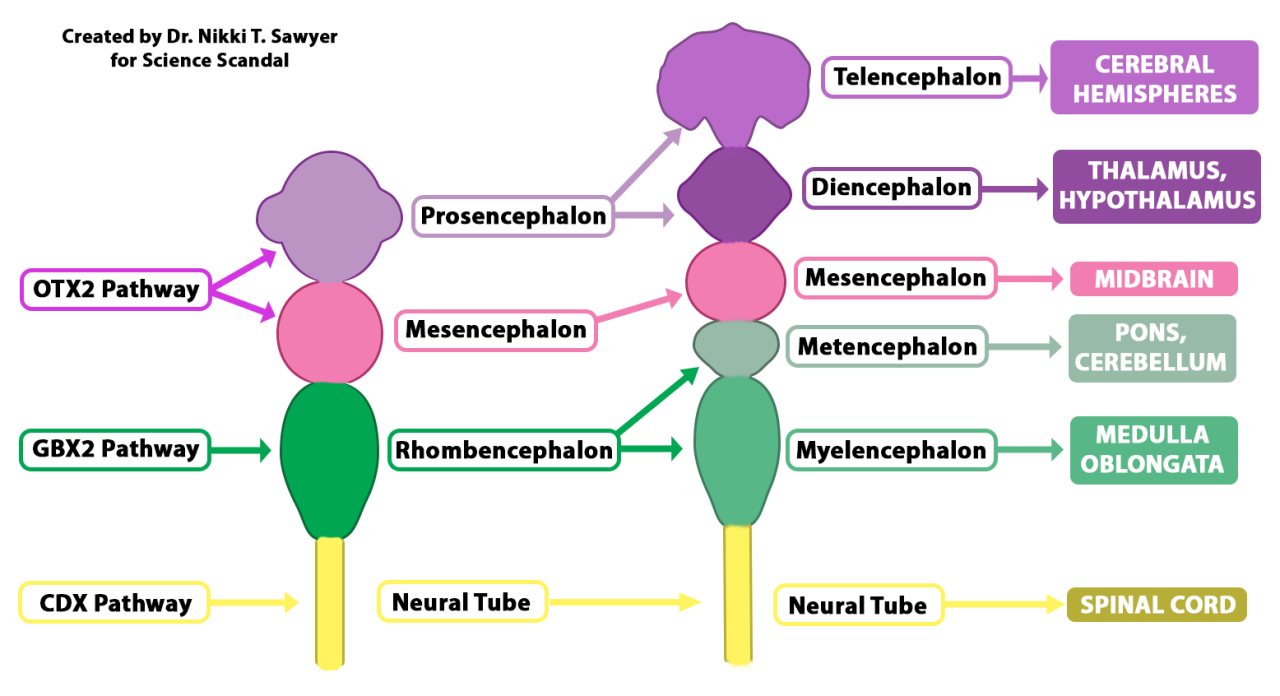
Traditionally, early brain development has been described as though the developing nervous system begins with a common population of neural precursor cells that is later divided into different regions. The familiar anatomy still follows the same sequence: the anterior neural tube forms the three primary brain vesicles, the prosencephalon, mesencephalon, and rhombencephalon, which later subdivide and develop into the major structures of the adult brain. Nothing in the new study changes that basic anatomical progression.
What the researchers found is that the developmental split appears to happen earlier than previously thought.
In simple terms, the early brain may begin with two different developmental pathways. During development, genes called homeobox genes help tell a cell, or group of cells, what developmental pathway to take. The researchers found that cells expressing the homeobox gene OTX2 give rise to the forebrain and midbrain, while a separate population expressing GBX2 gives rise to the hindbrain. When the researchers lineage-traced these cells in mouse embryos, the two populations remained separate rather than mixing together and later dividing into anterior and posterior brain regions.
The difference went deeper than simply turning on different gene names. The two populations also had different patterns of chromatin accessibility, meaning different parts of their DNA were available for use. Those molecular differences appeared to commit the cells to either a forebrain/midbrain fate or a hindbrain fate very early in development. When human pluripotent stem cells were directed into these two precursor states in the laboratory, they likewise showed different developmental potential.
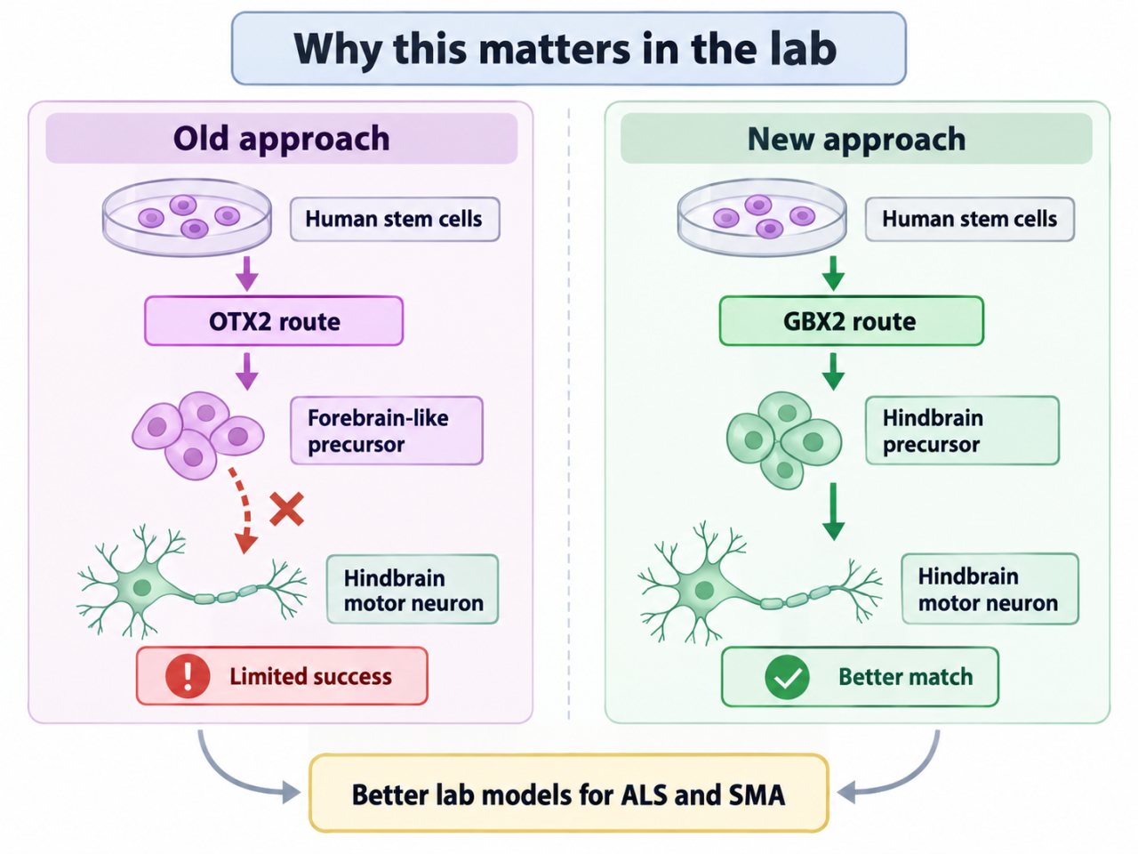
That finding helped solve a practical problem researchers had been struggling with for years. Scientists have been fairly successful at growing some types of forebrain neurons from human stem cells, but authentic hindbrain neurons have been considerably more difficult to produce. Once the researchers treated the hindbrain as arising through its own early developmental route rather than trying to push an anterior-type precursor toward a posterior fate, they were able to generate functional human motor neurons with characteristics of specific hindbrain regions. Those cells could eventually provide better laboratory models for diseases that affect these neurons, including spinal muscular atrophy and amyotrophic lateral sclerosis.
So that part is genuinely interesting.
But having different developmental pathways does not automatically make the resulting structure two separate organs.
Developmental biology routinely builds one organ from different populations of precursor cells following different molecular programs. The heart is a good example. Different early cardiac precursor populations make different contributions to the mature heart, including major contributions to different ventricles and the outflow tract. Those developmental differences are important for understanding both normal heart formation and congenital heart defects. Nevertheless, the finished structure is still one heart.
The brain study itself uses more careful language than many of the headlines. The authors say their results support the idea that the brain is a “composite organ” arising from two lineage-restricted progenitor populations. That is a developmental hypothesis about how one organ is assembled, not evidence that an adult human possesses two independently functioning brains that were physically fused together.
The researchers also found evidence that this anterior-versus-posterior developmental organization may be evolutionarily ancient. Related patterns were examined in zebrafish, chickens, and acorn worms, leading the authors to propose that the two developmental programs may extend back hundreds of millions of years. That evolutionary interpretation is intriguing, but it is broader than the central experimental result: the forebrain/midbrain and hindbrain appear to arise from distinct early neural progenitor populations.
And that is really the takeaway.
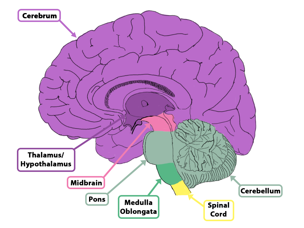
The discovery changes our understanding of the developmental ancestry of the hindbrain. It does not replace the three primary brain vesicles, rearrange the adult brain, or reveal another organ that anatomists somehow failed to notice.
Your brain is still one brain.
Its construction manual just turned out to have more than one set of instructions. 🧠
Sources
-
Two parallel neural ectoderm progenitors contribute to the developing brain
Jokhai, R. T., Dundes, C. E., Ahsan, H. S., et al. (2026). Two parallel neural ectoderm progenitors contribute to the developing brain. Nature Neuroscience.
Why this source matters: Primary source for the mouse lineage tracing; anterior versus posterior neural ectoderm; OTX2- and GBX2-associated progenitor populations; chromatin differences; human pluripotent-stem-cell differentiation; hindbrain motor-neuron generation; and the authors’ evolutionary/composite-organ hypothesis. The paper itself says the results support a two-progenitor model and that the authors postulate a composite-organ interpretation.
-
How to make a brain: new experiments challenge existing picture
Peeples, L. (2026, September 18). How to make a brain: new experiments challenge existing picture. Nature.
Why this source matters: Independent coverage useful for contextualizing how the findings are being interpreted outside the research team. It describes the evidence as challenging the common-progenitor model rather than presenting the discovery simply as “two brains.” It is also the source of Loh’s statement that the brain is one organ built from two different parts.
-
The human brain is two separate organs, study finds
Conger, K. (2026, September 18). The human brain is two separate organs, study finds. Stanford Medicine / Stanford Report.
Why this source matters: Useful both for explaining the research in accessible language and for documenting the article’s science-communication angle. Stanford’s own coverage explicitly escalates the paper’s “composite organ” hypothesis into the claim that the human brain consists of “two separate organs,” making it an important example of how the stronger wording entered the news cycle. It also summarizes OTX2/GBX2 lineage findings and the successful generation of hindbrain motor neurons.
-
The heart field transcriptional landscape at single-cell resolution
Kelly, R. G. (2023). The heart field transcriptional landscape at single-cell resolution. Developmental Cell, 58(4), 257–266.
Why this source matters: Review supporting the comparison that a single mature organ can be assembled from developmentally distinct precursor populations following different trajectories. It describes first- and second-heart-field progenitors contributing different regions to the definitive heart. This source supports the narrow point that multiple developmental programs do not, by themselves, establish the existence of multiple adult organs.