
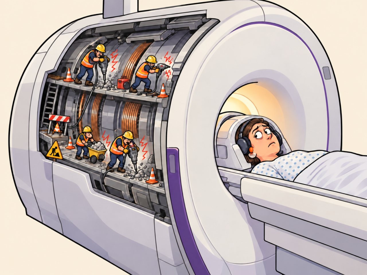
I recently had an MRI of my head, and while I was lying there in the machine, I started wondering. Because my mind can apparently never stop being curious.
I knew magnets didn’t make any noise. So what in the hell was causing all that loud banging and clanging around me?
If you’ve never had an MRI, you may not know that they usually give you BOTH earplugs AND headphones to protect your ears.
And you can still hear some variation of the following while lying in the tube:
BANG-BANG-BANG
THUNK… THUNK… THUNK
BRRRRRRRRRR
EEEEEEE-YOW-YOW-YOW
WEE-OO-WEE-OO-WEE-OO
Sometimes it even sounds like someone is summoning an angry dot-matrix printer trapped inside a washing machine.
And I’m lying there thinking, “Why does this sound like the machine is attacking me with a jackhammer?”
So, after coming home, I immediately jumped into research. I already knew the basics of how an MRI works, but where was all that noise coming from?
Turns out the main magnet itself is mostly innocent.
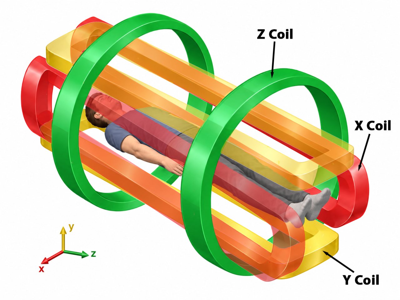
The real troublemakers are the gradient coils, which create small, controlled changes in the magnetic field along three different directions. Those gradients help the scanner determine where signals are coming from so the computer can reconstruct an image.
And get this: the currents in those gradient coils are switching rapidly while the coils sit inside the MRI’s enormous static magnetic field. That creates forces that make the coils and surrounding structures flex and vibrate.
Very tiny movements.
Absolutely ridiculous amounts of noise!
I also relearned that another set of coils, called RF coils, helps excite hydrogen nuclei and detect the signals used to build the image.
So, basically, what is happening is as follows:
Main magnet: “Everybody line up.” 🧲
RF coil: “Everybody dance!” 📡
Gradient coils: “YOU dance slightly differently depending on where you’re located.” 📍
Computer: “Excellent. I now know where everyone is.”
Gradient coils: BRRRRRRT THUNK THUNK THUNK THUNK
Me: “WHY DOES MEDICAL IMAGING SOUND LIKE ROAD CONSTRUCTION?” 🤣
The Real Story
An MRI scanner contains several different electromagnetic systems doing different jobs. The enormous main magnet creates a strong, nearly uniform magnetic field through the patient. That field influences hydrogen nuclei, especially the abundant hydrogen found in water and fat throughout the body. Radiofrequency, or RF, pulses then disturb those nuclei from their equilibrium state, and the scanner detects signals produced as they return toward equilibrium.
But detecting a signal is not enough. The scanner also has to figure out where inside the body that signal came from.
That is where the gradient coils enter the story.
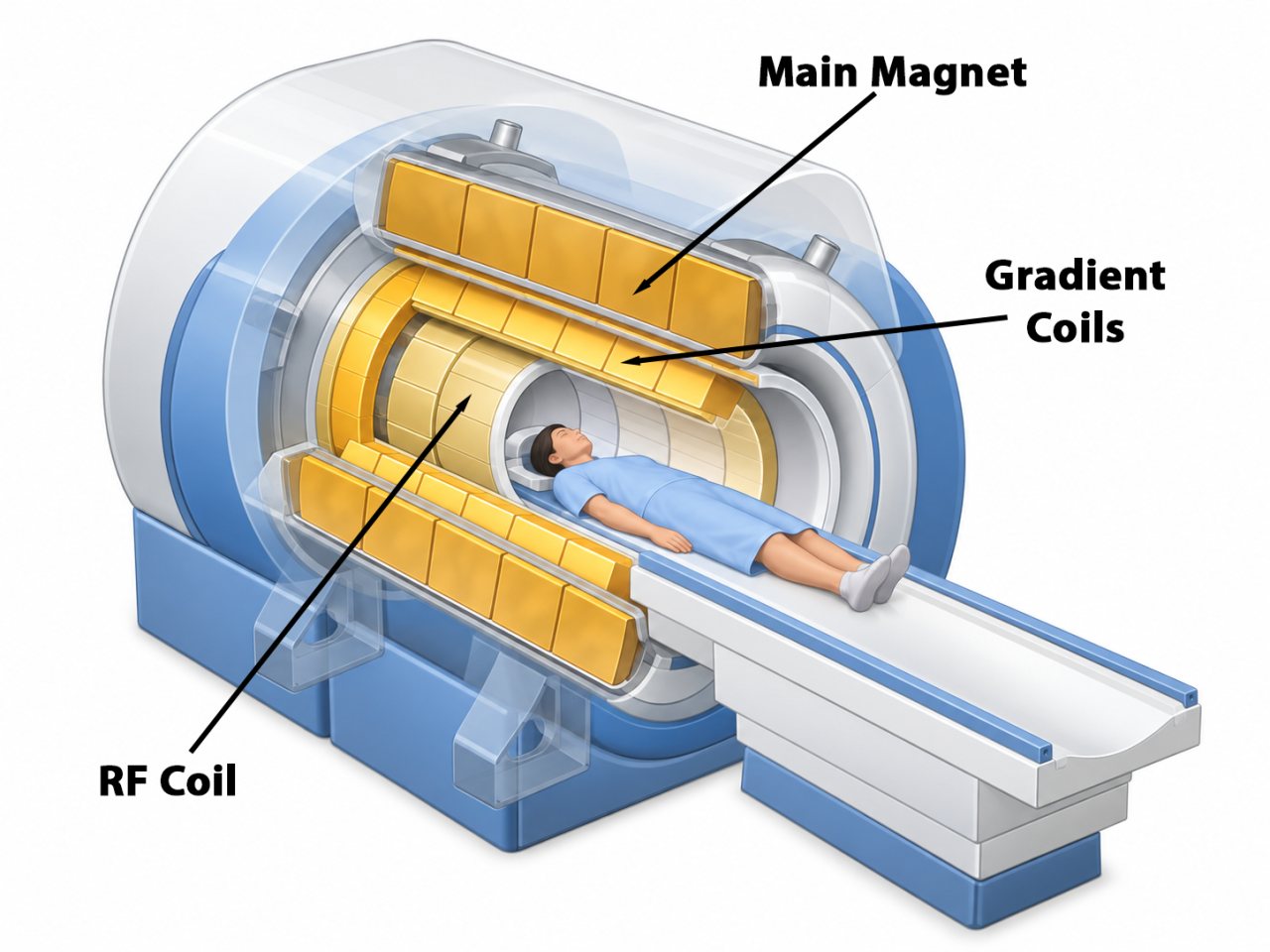
MRI scanners contain gradient coils that create small, carefully controlled variations in the magnetic field across three spatial directions. Because the resonant behavior of hydrogen depends on the local magnetic field, those gradients allow the scanner to encode position into the signals it receives. In simplified terms, the gradient system gives the MRI a way to distinguish “signal coming from over here” from “signal coming from over there.”
Unfortunately for everyone’s ears, doing that quickly requires rapidly changing large electrical currents through the gradient coils.
Those current-carrying conductors are sitting inside the MRI scanner’s extremely strong static magnetic field. Physics therefore supplies a Lorentz force: a force on an electrical current moving through a magnetic field. As the gradient currents rapidly change in strength and direction, the forces acting on the gradient-coil conductors change too.
The conductors and their supporting structures respond by vibrating.
The hardware barely has to move at all. Tiny vibrations inside a rigid structure can produce an acoustic tantrum loud enough to require earplugs.
Those vibrations travel through the gradient assembly and other parts of the scanner and ultimately push on the surrounding air, producing the bangs, knocks, buzzes, hums, and alarming mechanical noises heard by the patient.
So the giant magnet itself is not sitting there going BANG-BANG-BANG.
The noise is largely the mechanical consequence of rapidly switching gradient currents inside that magnetic field.
And this also explains why an MRI does not make just ONE characteristic sound.
Different MRI pulse sequences use the gradients in different patterns, with different amplitudes, timing, switching rates, and combinations of axes. Change the electrical pattern driving the gradients, and you change the forces and vibrations produced by the scanner. Different vibration frequencies can also interact differently with the mechanical resonances of the scanner structure.
That is why one part of an examination may sound like:
THUNK… THUNK… THUNK
while the next abruptly becomes:
BRRRRRRRRRR
followed by something apparently designed by an engineer who had unresolved feelings about dot-matrix printers.
The noise is not merely annoying. Some MRI sequences can produce sound-pressure levels above 100 decibels. Echo-planar imaging, a particularly gradient-intensive technique widely used in functional and diffusion MRI, can reach roughly 110–120 decibels in some systems. At those levels, hearing protection is not just a courtesy. It is a safety measure.
That is why patients may be given earplugs, headphones, or both.
Engineers have developed quieter gradient designs, vibration isolation, sound-absorbing materials, active noise-control systems, and imaging sequences that reduce rapid gradient switching. Some newer “quiet” or “silent” MRI techniques can dramatically reduce the acoustic racket.
But in a conventional scanner, the explanation is wonderfully disproportionate:
The machine needs extremely precise magnetic-field changes to determine where signals originated.
Producing those changes requires rapidly switching powerful electrical currents.
Those currents experience forces inside the main magnetic field.
The scanner vibrates.
And suddenly microscopic anatomy is being mapped while the patient listens to what sounds like road construction occurring six inches from their skull.
Sources
-
Overview of Methods for Noise and Heat Reduction in MRI Gradient Coils
Motovilova, E., & Winkler, S. A. (2022). Overview of methods for noise and heat reduction in MRI gradient coils. Frontiers in Physics, 10, 907619.
Why this source matters: Explains the central mechanism behind MRI acoustic noise: rapidly switched currents in gradient coils experience Lorentz forces within the scanner’s static magnetic field, causing mechanical vibration. It also discusses gradient-related noise levels, pulse-sequence effects, and engineering approaches used to reduce MRI noise.
-
Magnetic Resonance Imaging (MRI)
National Institute of Biomedical Imaging and Bioengineering. Magnetic Resonance Imaging (MRI). National Institutes of Health.
Why this source matters: Provides an authoritative overview of how MRI uses strong magnetic fields, hydrogen protons, radiofrequency excitation, and detected signals to create images. It also notes that MRI scanners can produce very high sound levels and that patients receive hearing protection.
-
Benefits and Risks: MRI
U.S. Food and Drug Administration. Benefits and Risks: Magnetic Resonance Imaging (MRI). Last updated 12/09/2017.
Why this source matters: Provides the patient-safety side of MRI acoustic noise. The FDA identifies the loud knocking sounds produced during scanning as a potential hearing hazard and explains the need for appropriate hearing protection.
-
Acoustic Noise Concerns in Functional Magnetic Resonance Imaging
Moelker, A., & Pattynama, P. M. T. (2003). Acoustic noise concerns in functional magnetic resonance imaging. Human Brain Mapping, 20(3), 123–141.
Why this source matters: Provides a detailed explanation of how fluctuating gradient currents create Lorentz forces that bend and vibrate gradient coils and related structures, with those vibrations propagating through the scanner and becoming airborne sound. It also discusses how MRI acoustic noise varies with gradient waveforms and pulse sequences.
-
Measuring the vibrational response of a high performance gradient coil
Kelley, D. A. C. (2026). Measuring the vibrational response of a high performance gradient coil. Magnetic Resonance Imaging, 134, 110766.
Why this source matters: This technical study directly examines vibration in a high-performance MRI gradient coil. It explains that gradient coils are deliberately engineered to be very rigid and massive to minimize motion, yet small residual vibrations still occur and are associated with the acoustic noise generated by the coil. The study experimentally measures the gradient system’s mechanical response and relates those vibrations to the structure and mounting of the coil.