
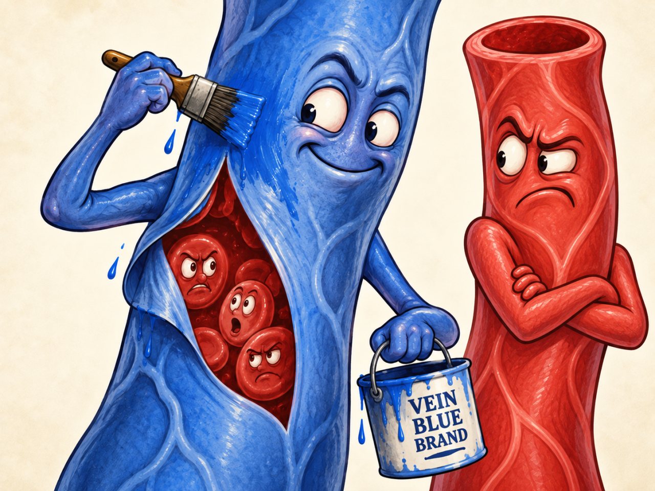
As someone who taught Anatomy & Physiology for many semesters, I got very used to students arriving with one particular piece of anatomical misinformation already firmly installed:
There is red blood. And there is blue blood.
Honestly, I can’t entirely blame them.
Open almost any anatomy textbook and the systemic arteries are blazing red while the systemic veins are painted a very convincing shade of blue. Anatomical models do the same thing.
It looks official.
It is also absolutely not the color of the blood inside those veins.
And while we’re cleaning up circulatory propaganda, there’s another misconception hiding underneath it:
Arteries are not “the red vessels,” and veins are not “the blue vessels.”
Arteries carry blood away from the heart. Veins carry blood back toward the heart.
Or, as A&P instructors like to reduce several paragraphs of physiology to four words:
Arteries supply. Veins drain.
Most systemic arteries happen to carry oxygen-rich blood, so we color them red. Most systemic veins happen to carry oxygen-poor blood, so we color them blue.
But pulmonary circulation and fetal circulation stroll in and ruin the color scheme immediately.
Pulmonary arteries and umbilical arteries carry oxygen-poor blood.
Pulmonary veins and the umbilical vein carry oxygen-rich blood.
So where did humans get the idea that venous blood itself is blue?
Unfortunately, your own body is helping spread the rumor.
Look at the superficial veins beneath pale skin and they really can appear blue, blue-green, or purple.
But cut one open and you will not uncover a tiny plumbing system full of blueberry juice.
The blood inside is red.
It has always been red.
Your skin has just been running an extremely convincing optical scam.
So, to summarize the three-layer anatomy scam:
Misconception #1: Blood can be blue.
Nope. Deoxygenated human blood is dark red.
Misconception #2: Arteries are red and veins are blue.
Nope. Those colors usually indicate oxygenation, not vessel type.
Misconception #3: Pulmonary vessels are the only “exceptions” to the color rule.
Nope again. Fetal circulation joins the rebellion. Umbilical arteries carry oxygen-poor blood, while the umbilical vein carries oxygen-rich blood.
The Real Story
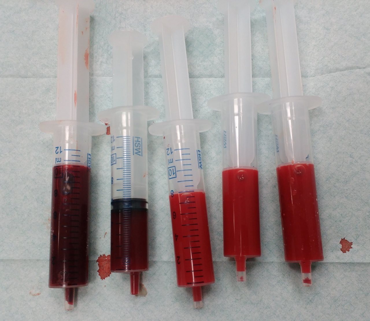
Human blood is always some shade of red.
The color comes largely from hemoglobin, the oxygen-carrying protein inside red blood cells. Each hemoglobin molecule contains iron-bearing heme groups that bind oxygen. When hemoglobin is carrying more oxygen, blood appears brighter red. After oxygen is released to the tissues, deoxygenated hemoglobin gives venous blood a darker, deeper red. It does not turn blue.
So why can superficial veins look blue through the skin?
The answer is more complicated than the familiar explanation that “blue light scatters more.”
Light entering the skin is absorbed and scattered differently depending on its wavelength. Red light penetrates tissue more deeply than blue light, so a vein beneath the skin affects the returning red light more strongly. Blood absorbs some of that light, and the skin above a vein therefore returns a different mixture of wavelengths to the eye than the surrounding skin does. Vessel depth and diameter, blood oxygenation, skin optics, and our own visual perception all contribute to the final color we see.
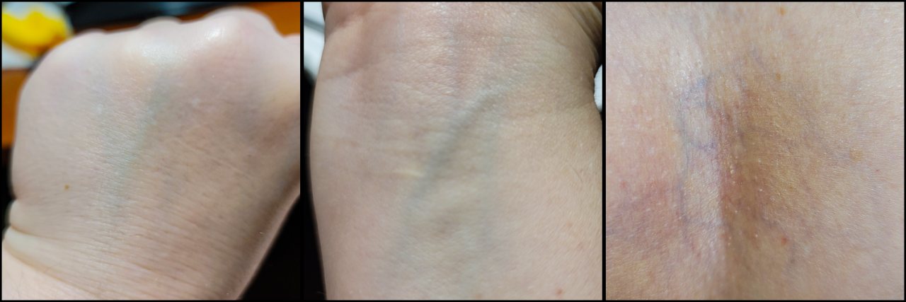
In fact, the classic experimental study of blue-looking veins found that there is no single “blue pigment” or simple blue-reflection trick responsible. The apparent color emerges from the interaction of blood, tissue, light, and the visual system. Later work has also shown that scattering by structures within the dermis can contribute to the effect.
That also explains why veins do not look equally blue on everyone. Their appearance can change with skin pigmentation, vessel depth, vessel size, lighting, surrounding skin color, and oxygenation. Some may look blue, others blue-green or purple, and some may barely be visible at all. One perception study even found that the apparent blueness became stronger when the vein was viewed against the surrounding skin, showing that color contrast in the brain also helps create the effect.
And then there is cyanosis.
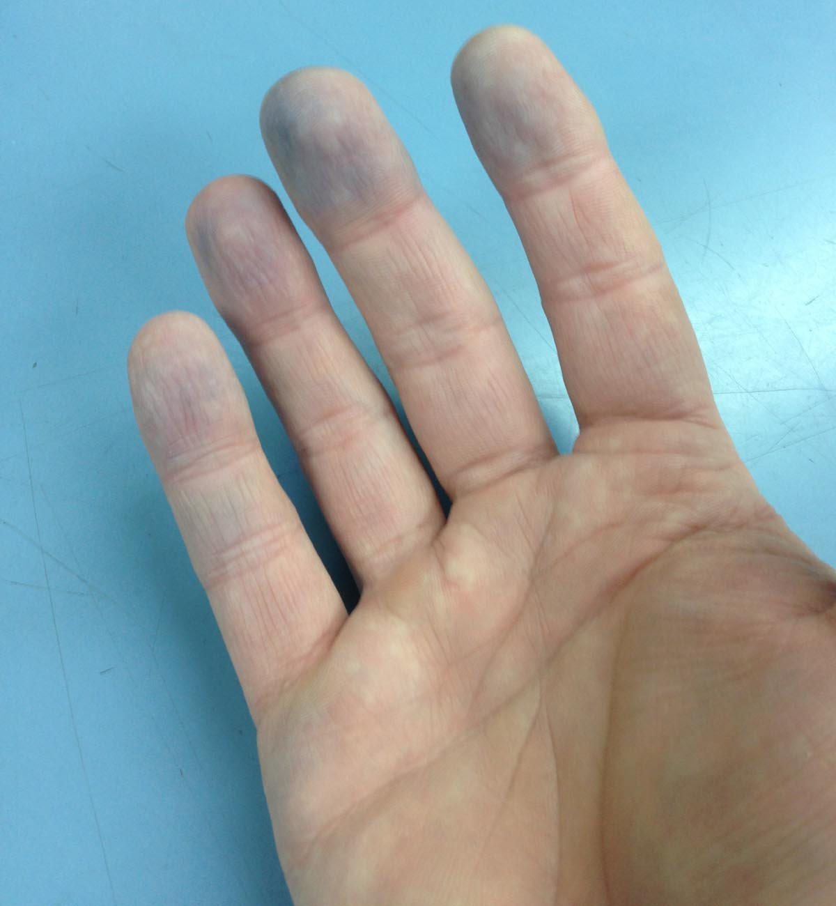
Cyanosis is a bluish or grayish discoloration of the skin or mucous membranes that can occur when the amount of deoxygenated hemoglobin becomes unusually high. But even then, the blood itself has not turned blue. Reduced oxygenation makes hemoglobin, and therefore the blood, darker red. Viewed through tissue, that change in optical properties can contribute to a blue or bluish-purple appearance at the surface.
The red-and-blue convention used in anatomy diagrams is therefore exactly that: a convention.
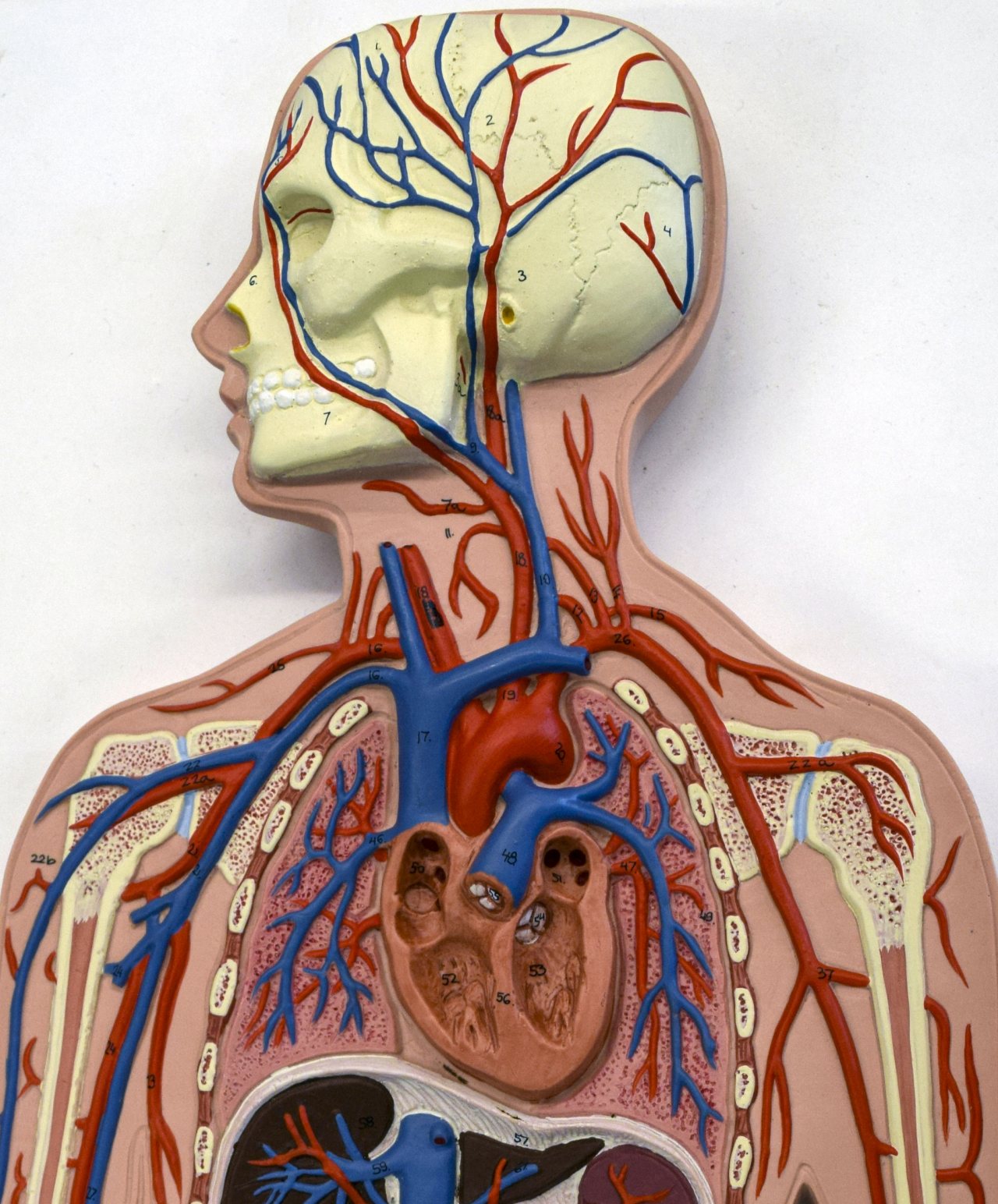
Arteries are defined by carrying blood away from the heart, and veins by carrying blood towards the heart. In the systemic circulation, arteries usually carry oxygen-rich blood and veins usually carry oxygen-poor blood. In the pulmonary circulation, that pattern reverses: pulmonary arteries carry oxygen-poor blood to the lungs, while pulmonary veins return oxygen-rich blood to the heart.
Fetal circulation adds another reminder not to confuse vessel type with oxygen content. The two umbilical arteries carry relatively oxygen-poor blood from the fetus toward the placenta, while the single umbilical vein carries oxygen-rich blood from the placenta back toward the fetus.
So the textbooks are not claiming that venous blood is blue.
They are simply using blue ink to represent lower oxygen levels.
Your actual circulatory system declined to adhere to the textbook color scheme.
Sources
-
Why do veins appear blue? A new look at an old question
Kienle A, Lilge L, Vitkin IA, Patterson MS, Wilson BC, Hibst R, Steiner R. “Why do veins appear blue? A new look at an old question.” Applied Optics. 1996;35(7):1151–1160.
Why this source matters: This is the key primary optics paper. It identifies skin absorption/scattering, blood oxygenation, vessel depth and diameter, and visual perception as contributors to apparent vein color.
-
18.1 An Overview of Blood
Betts JG, Young KA, Wise JA, et al. Anatomy and Physiology 2e. Houston, TX: OpenStax; 2022. Section 18.1, “An Overview of Blood.”
Why this source matters: Supports the distinction between bright-red oxygenated blood and darker-red blood after oxygen release.
-
39.4 Transport of Gases in Human Bodily Fluids
Clark MA, Douglas M, Choi J. OpenStax. Biology 2e. Section 39.4, “Transport of Gases in Human Bodily Fluids.”
Why this source matters: Explicitly states that the heme group contains iron, oxygenated blood is brighter red, and deoxygenated venous blood is darker red.
-
Cyanosis
Pahal P, Goyal A. Cyanosis. In: StatPearls [Internet]. Treasure Island (FL): StatPearls Publishing. Updated October 3, 2022.
Why this source matters: Useful for the cyanosis section because it explicitly describes increased deoxyhemoglobin, darker-red blood, and bluish surface appearance. It also supports the clinical distinction between cyanosis as a visible discoloration and actual blood color.
-
20.1 Structure and Function of Blood Vessels
Betts JG, Young KA, Wise JA, et al. Anatomy and Physiology 2e. Houston, TX: OpenStax; 2022. Section 20.1, “Structure and Function of Blood Vessels.”
Why this source matters: Explicitly defines arteries as carrying blood away from the heart and veins as carrying blood toward it, and distinguishes oxygen content in systemic versus pulmonary vessels.
-
Fetal Circulation
American Heart Association. “Fetal Circulation.” American Heart Association. Last reviewed June 10, 2026.
Why this source matters: Confirms that oxygen-rich blood travels from placenta to fetus through the umbilical vein, while blood returns to the placenta through the umbilical arteries.